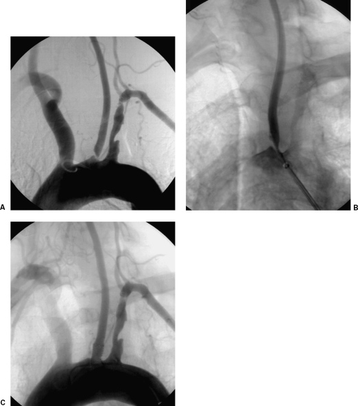
Figure 2.
Interventional treatment of a left common carotid artery (CCA) ostial lesion. (A) Arch digital subtraction angiography (DSA) using a pigtail catheter depicts a high-grade left CCA ostial lesion. (B) DSA via a long sheath placed in the aortic arch is used to place the stent accurately; note the guidewire already placed across the lesion. (C) Completion DSA depicts successful recanalization with the use of a balloon-expandable stent. (Images courtesy of Bryan T. Petersen, M.D., Dotter Interventional Institute, Health and Sciences University, Oregon.)

