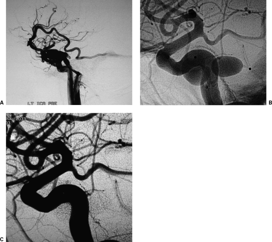
Figure 8.
Detachable balloon occlusion technique for the treatment of a traumatic carotid cavernous fistula (TCCF) in a patient with diplopia and headaches following head injury. (A) Lateral projection of left internal carotid artery (ICA) digital subtraction angiography (DSA) depicts the TCCF. The laceration is located at the C5 segment. (B) Nonsubtracted images depict two detachable balloons deployed into the left cavernous sinus. (C) Completion DSA depicts complete sealing of the TCCF with good ICA flow.

