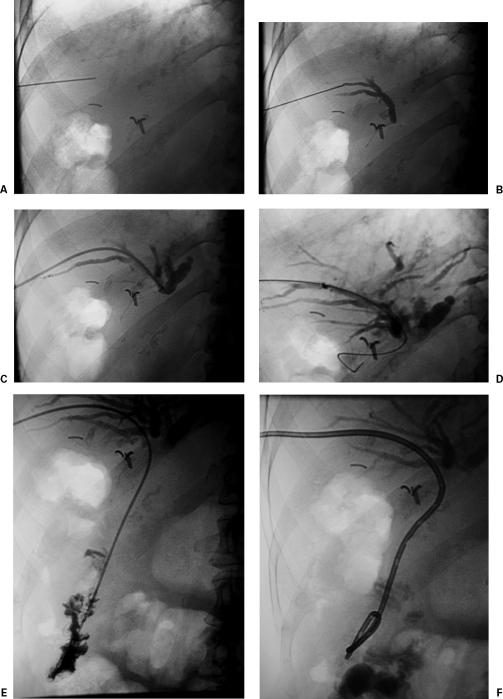
Figure 1.
Percutaneous transhepatic biliary drainage in a 71-year-old woman with pancreatic carcinoma. (A) Fluoroscopic image shows needle being retracted in the liver after initial puncture. (B) Fluoroscopic image shows peripheral bile duct opacification. (C) Fluoroscopic image demonstrates 6F outer dilator of the AccuStick kit (Boston Scientific, Natick, MA) in the central bile ducts. (D) Fluoroscopic image shows 5F Kumpe (Cook, Bloomington, IN) catheter and guidewire advanced across the site of obstruction to expected region of small bowel. (E) Fluoroscopic image shows the 5F Kumpe catheter has been advanced into the small bowel. (F) Final fluoroscopic image shows 8F biliary drainage catheter in good position.

