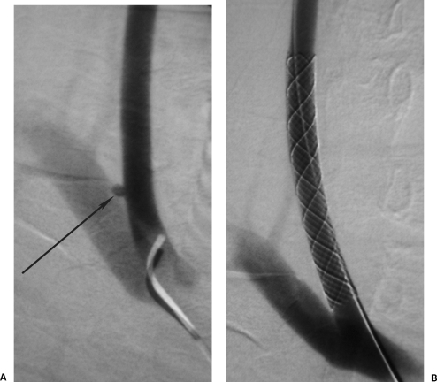
Figure 12.
Low knife wound to the right neck with a large hematoma and initial loss of consciousness. (A) Digital subtraction angiography of the common carotid artery shows a small proximal common carotid pseudoaneurysm with no evidence of dissection. (B) Follow-up carotid angiogram shows exclusion of the false aneurysm by a Wallgraft (Boston Scientific, Natick, MA). (Images courtesy of Dr. David Kessel.)

