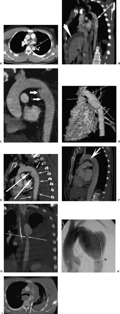
Figure 2.
The spectrum of aortic injuries. (A) Axial computed tomography (CT) and (B) sagittal oblique reformat of focal intimal flap (thin arrow, intimal flap; thick arrow, perihepatic hematoma from liver contusion). (C) CT reconstruction demonstrates a focal aortic injury at the isthmus. (D) A surface rendered CT image shows a circumferential aortic injury. (E) A 47-year-old man crushed by a steel door. CT reformat shows focal aortic injury (large arrows) with extensive intramural hematoma (small arrows). (F) A 5-year follow-up CT of a post-traumatic dissection shows the entry tear distal to the left subclavian artery (arrow). Minimal false lumen expansion had occurred over this period. (G) Coronal CT reformat shows pseudocoarctation. (H) A 36-year-old with a 6.5-cm late saccular aneurysm. The patient had had a polytrauma motorbike accident 18 years previously. A thoracic angiogram shows an aneurysm that was incidentally detected on a diving medical chest radiograph. (I) A 24-year-old male car driver. Axial CT scan shows active bleeding (arrows) from an aortic injury. The patient had a lethal head injury and died within 1 hour of this scan.

