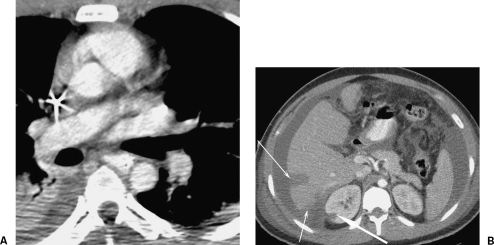
Figure 4.
A complication of an intimal flap. A 27-year-old construction worker fell 15 feet onto a scaffolding bar. His initial aortic images are shown in Fig. 2A and B. The thickness of the flap suggests there may be thrombus formation on it. His life-threatening liver contusion and active hemorrhage on computed tomography (CT) was successfully treated by coil embolization (not shown). Follow-up axial CT examination 7 days later (A) at the same level as Fig. 2A show no evidence of an intimal flap and (B) a new right renal infarction (broad arrow). This became a focal scar on follow-up. The hepatic laceration is shown (narrow arrows). If the embolus had passed into one of the mesenteric vessels, it may have had serious consequences.

