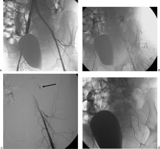
Figure 3.
Angiogram with embolization of left inferior epigastric artery. (A) Pelvic arteriogram shows normal appearing arteries without evidence of active extravasation. Note the bladder deviated to the left. (B) Arterial phase of a selective left common iliac angiogram shows active bleeding from the inferior epigastric artery. (C) Digital subtraction left common iliac angiogram shows bleeding site (arrow). (D) Inferior epigastric angiogram after coil embolization shows complete occlusion of vessel.

