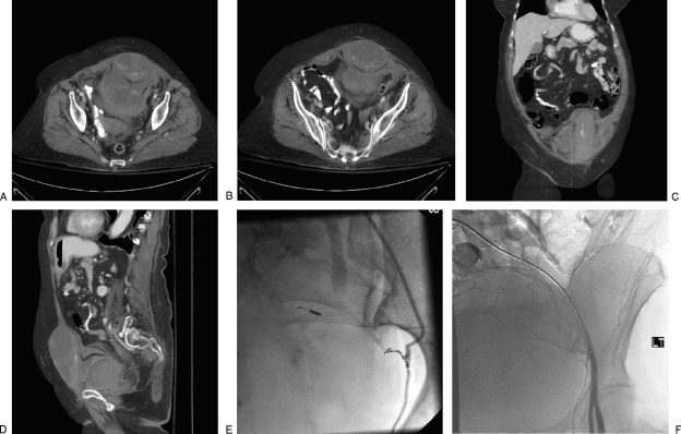
Figure 5.
Spontaneous rectus sheath hemorrhage in an anticoagulated patient. (A, B) Axial enhanced computed tomography (CT) images show large rectus sheath hematoma ruptured into the prevesical space with active bleeding. (C) Coronal reformatted CT image shows bleeding site in left rectus muscle. (D) Sagittal reformatted CT image shows extent of hemorrhage in prevesical space. (E) Angiogram of left inferior epigastric artery shows vasospasm of the artery. (F) Angiogram after embolization shows complete occlusion of the artery.

