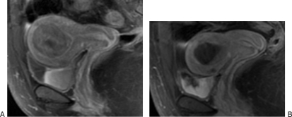
Figure 1.
(A) A 44-year-old African American woman with history of menorrhagia, pelvic pain, and urinary frequency. Saggital T1 magnetic resonance imaging (MRI) with contrast shows a diffusely enhancing intramural fibroid that measures 5.5 × 4.2 cm with associated mass effect on the bladder and displacement of the endometrial cavity. There is also a smaller anterior intramural fibroid. (B) At 6-month follow-up, MRI after uterine artery embolization (UAE) demonstrates complete infarction of the fibroid with significant decrease in size of the tumor, which now measures 2.7 × 2.1 cm. There is normal enhancement of myometrium. The patient was symptom free at follow-up.

