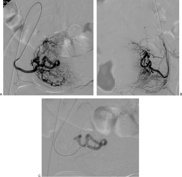
Figure 4.
(A) Same patient as in Fig. 2. Selective oblique view angiogram of the right uterine artery after forming a Waltman loop with an end-hole catheter from an ipsilateral puncture. There is identification of the cervicovaginal branch. (B) Contralateral left uterine artery angiogram. (C) Postembolization angiogram after introduction of a microcatheter that was advanced beyond cervicovaginal branch. Embolization was performed using 355 to 500 μm nonspherical polyvinyl alcohol. Angiogram shows stasis of the contrast column for several heartbeats and no further enhancement of fibroid mass.

