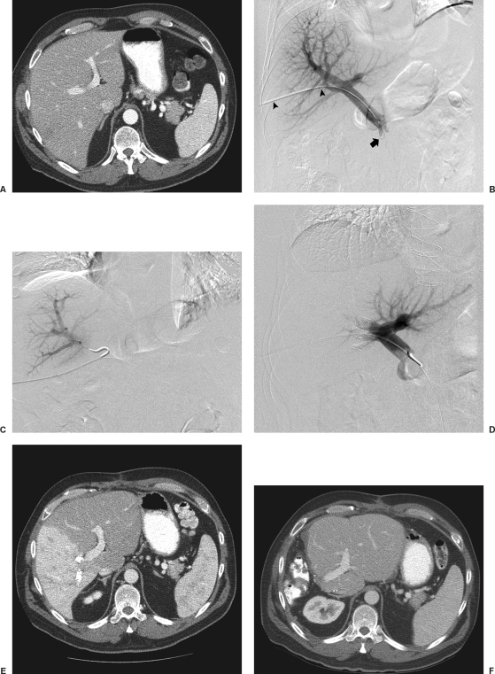
Figure 1.
Transhepatic ipsilateral right PVE using tris-acryl particles and coils performed in a 49-year-old with colon cancer metastatic to the liver. (A) Computed tomography (CT) scan obtained prior to portal vein embolization (PVE) shows marginal future liver remnant (FLR) (FLR-to-TELV (total estimated liver volume) ratio = 25%). (B) Anteroposterior flush portogram shows a 6F vascular sheath in a right portal vein branch (arrowheads) and a 5F flush catheter within the main portal vein (arrow). (C) Selective right portogram with use of reverse-curve catheter during right PVE. (D) Final portogram shows occlusion of the portal vein branches to segments V through VIII with continued patency of the veins supplying the left lateral lobe. (E) CT scan obtained 1 month after right PVE shows substantial FLR hypertrophy (FLR-to-TELV ratio = 50%). The degree of hypertrophy is 25%. (F) CT scan after right hepatectomy shows hypertrophy of remnant liver.

