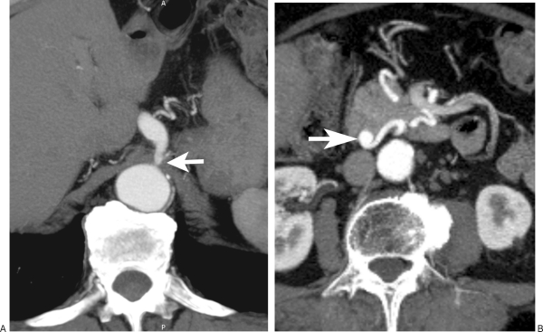
Figure 6.
Incidentally detected small inferior pancreaticoduodenal artery aneurysm in a 50-year-old man. (A) Axial subvolume (4.75 mm thick) maximum intensity projection (MIP) of a computed tomography (CT) angiography dataset shows a high-grade stenosis (arrow) at the origin of the celiac axis. (B) Axial subvolume (4.75 mm thick) MIP of a CT angiography dataset shows a small aneurysm (arrow) within the head of the pancreas.

