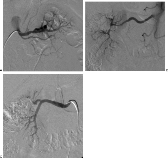
Figure 3.
(A) Angiographic appearance of segmental arterial mediolysis (SAM). Catheter angiography demonstrates segmental alternating fusiform and saccular appearance of the distal left renal artery, representing dissecting aneurysms. (B) Angiographic appearance of SAM. Catheter angiography demonstrates segmental stenoses and irregularity of the distal right renal artery with resultant occlusion of the interlobar and interlobular arteries. (C) Angiographic appearance of SAM. Catherter angiography demonstrates fusiform enlargement of the right renal artery, representings a dissecting aneurysm.

