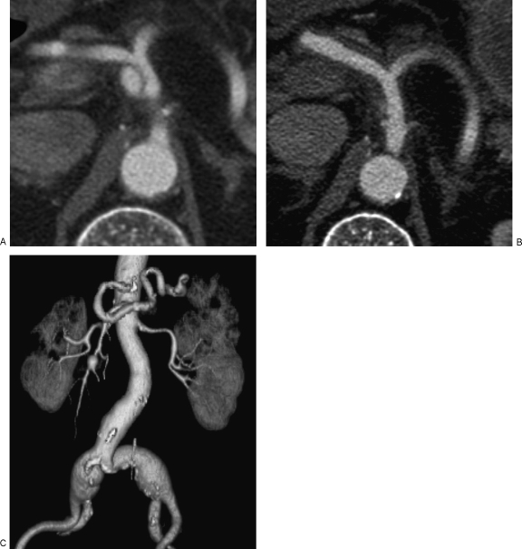
Figure 5.
(A) Dissecting hematomas in segmental arterial mediolysis (SAM). Axial contrast-enhanced computed tomography (CT) scan demonstrates a dissecting aneurysm involving the celiac artery. (B) Dissecting hematomas in SAM. Axial contrast-enhanced CT scan is more sensitive for depiction of diffuse arterial wall thickening in the common hepatic and splenic arteries representing dissecting hematomas. (C) Dissecting hematomas in SAM. Coronal three-dimensional reconstructed image in the same patient demonstrates additional saccular aneurysms in the splenic and superior mesenteric arteries as well as fusiform aneurysms involving the bilateral common iliac arteries.

