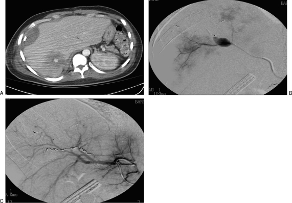
Figure 4.
(A) A computed tomography (CT) scan of the abdomen demonstrating multiple foci of septic emboli, in a patient with bacterial endocarditis. (B) On angiography, note is made of an infectious pseudoaneurysm from the third-order branches of the right hepatic artery. (C) This was treated with transcatheter coil embolization.

