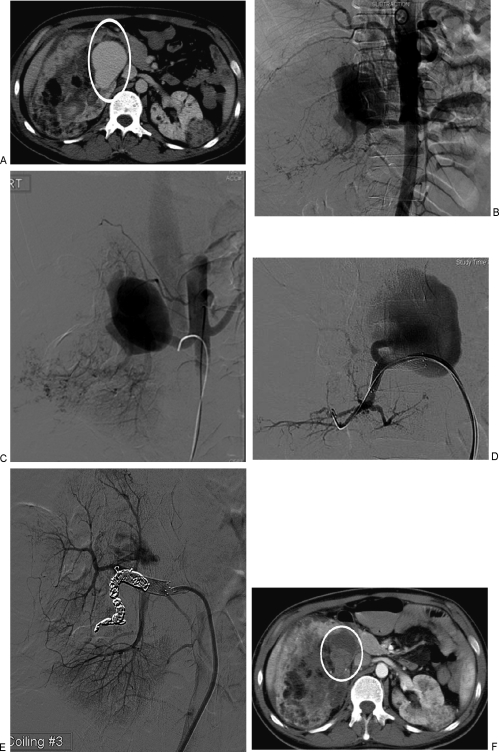
Figure 7.
Renal arteriovenous malformation (AVM). (A) Axial computed tomography (CT) scan with bilateral angiomyolipoma in a patient with tuberous sclerosis. Area in white ellipse shows the AVM in the right kidney. (B) Aortogram demonstrates the right renal AVM and classic angiographic findings of angiomyolipoma in the lower right renal arterial branches. (C) Selective right renal arteriogram demonstrates the AVM and early filling of the inferior vena cava. (D) Super-selective arteriogram confirms a single feeder into the AVM from a segmental renal artery branch. A stent has been placed in the main renal artery to assist in coiling of the feeder. (E) Successful coil embolization of the feeder to the AVM. Note minimal residual flow in the AVM. (F) Follow-up CT the next day demonstrates complete thrombosis of the AVM (in white circle).

