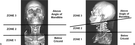
Figure 1.
Zones of the neck. Anterior (A) and lateral (B) views of a 3-D reconstruction of a computed tomographic angiogram of the neck demonstrate the three anatomic zones. Zone I extends from the thoracic inlet up to the inferior border of the cricoid cartilage. Zone II extends from the cricoid cartilage to the angle of the mandible. Zone III extends from the angle of the mandible up to the skull base. Injured vessels within zones I and III may be very difficult to identify and control, and may require an arteriogram to evaluate and plan further interventions.

