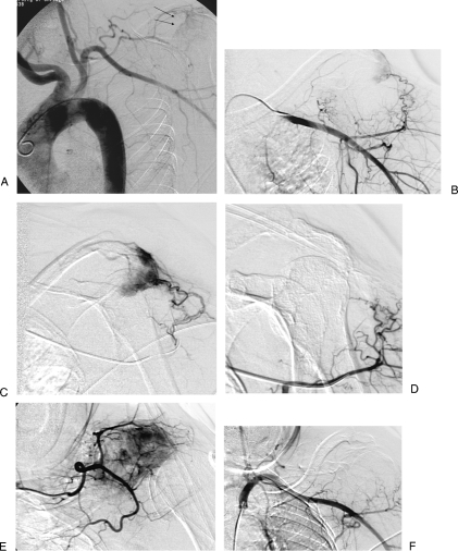
Figure 4.
A 68-year-old woman with renal cell carcinoma metastasis to left scapula. (A) Aortogram shows tumoral blush of right scapula (black arrows). (B) Selective axillary angiogram shows tumoral vessels. (C) Superselective angiogram through microcatheter shows tumor. (D) Superselective angiogram after embolization with trisacryl beads shows occlusion of tumoral vessels. (E) Superselective angiogram of second tumoral feeding vessel shows tumoral blush. (F) Final subclavian angiogram after additional particle embolization shows occlusion of tumoral vessels.

