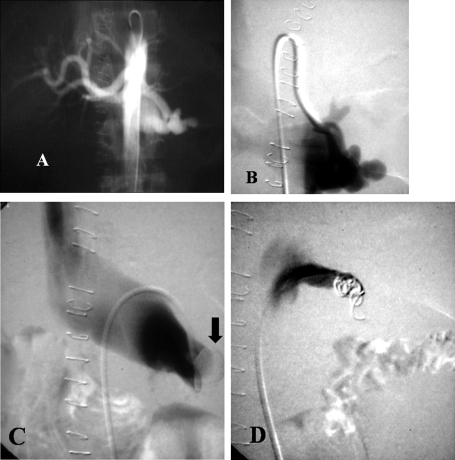
Figure 6.
Patient with a large left renal artery arteriovenous fistula (AVF) after a nephrectomy. (A) Angiogram shows an AVF of the stump of the left renal artery. (B) Selective left renal digital subtraction angiogram shows rapid filling of the left renal vein. (C) Digital subtraction angiogram venogram shows an occlusion balloon placed in the left renal vein to decrease flow and prevent coil migration. (D) Digital subtraction angiogram after embolization shows successful exclusion of the AVF.

