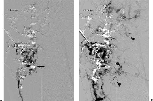
Figure 14.
Venous malformation (VM) sclerotherapy using opacified sclerosant and digital subtraction angiography technique. (A) DSA mask obtained after opacification of a large dysmorphic type II to III VM reveals initial radiolucent sclerosant (arrow) displacing contrast. (B) Addition of ethiodized oil to ethanol allows better opacification of agent within the periphery of lesion (arrowheads) and increased viscosity and therefore “dwell-time” of agent.

