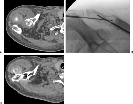
Figure 5.
Painful metastasis of the clavicle treated with cryoablation. (A) Computed tomography (CT) scan shows the osteolytic lesion with pathological fracture. (B, C) Two crossed cryoprobes are inserted coaxially through bone trocars to adapt the shape of the ice ball to the shape of the tumor. The hypodense ice ball is precisely monitored with CT (C). No further consolidation is required in this case.

