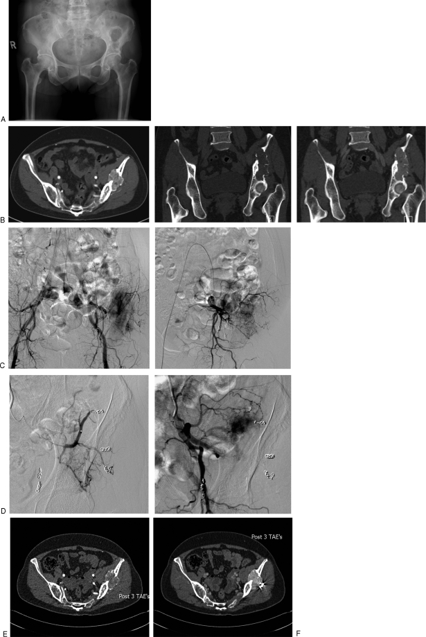
Figure 3.
Transarterial embolization (TAE) in a functional phosphaturic mesenchymal tumor causing oncogenic osteomalacia. The patient presented with pelvic pain with a 10-year history of a lytic lesion in the pelvis (A) thought originally to represent a brown tumor. Three separate embolization procedures were performed. Temporary improvement in fibroblast growth factor 23 occurred, but the tumor rapidly revascularized. (B) Coronal computed tomography (CT) pre- and postcontrast and axial contrast-enhanced CT demonstrating the hypervascular lesion in the left ileum extending to the acetabular surface. (C) Second embolization visit: flush aortogram demonstrates multiple coils in the superior gluteal artery with persistent arterial blush in the ileum; selective internal iliac angiogram demonstrates remaining arterial feeders. (D) Superior gluteal artery embolization: superior gluteal artery is occluded, with residual tumor blush from unnamed ileal artery. (E,F) CT 6 weeks after third TAE. Early arterial image demonstrated reduced vascularity but this fills in in late arterial phase (F).

