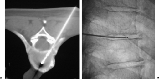
Figure 10.
Percutaneous thoracic radiofrequency nucleotomy. (A) The electrode in inserted into the disc, parallel to the adjacent vertebral end plates, between the head of the rib and the pedicle. (B) Lateral fluoroscopic view shows the tip of the electrode, away from the vertebral end plates. Due to reduced height of thoracic discs, only lateral Coblation® channels are dug to avoid damage to the vertebral end plates.

