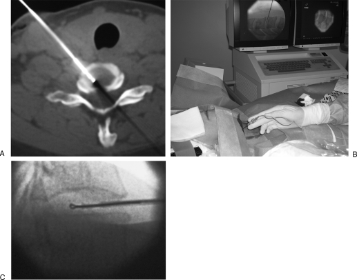
Figure 11.
Percutaneous cervical radiofrequency nucleotomy. (A, B) A dedicated 19-gauge radiofrequency electrode is inserted into the cervical disc via an anterolateral approach, just medial to the carotid artery. (C) Three spherical voids are created in the disc by rotation of the looped-tip radiofrequency electrode in the nucleus. The electrode should not be advanced beyond the midthird/posterior-third junction of the disc to avoid damage to the neurological structures.

