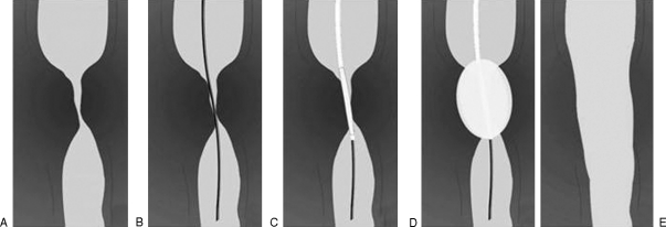
Figure 1.
Steps in fluoroscopically guided balloon dilation. (A) Stricture is identified on the preprocedural esophagogram. (B) Guide wire is advanced under fluoroscopic control across the lesion. (C) Balloon catheter is advanced over the wire and balloon is centered on the lesion. (D) Balloon is inflated, repeatedly if necessary. (E) Postprocedural esophagogram is performed to evaluate for esophageal perforation.

