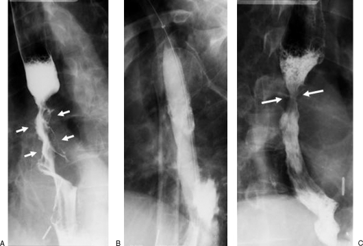
Figure 3.
A 68-year-old man with esophageal carcinoma. (A) Preprocedural esophagogram demonstrates a long irregular stricture in the midthoracic esophagus with shouldered margins caused by the tumor (arrows). (B) Under careful fluoroscopic control, the balloon is inflated to its maximum diameter of 20 mm. (C) Postprocedural esophagogram shows marked improvement of the stricture with an area of residual narrowing at the proximal end of the tumor (between arrows). There is no evidence of perforation.

