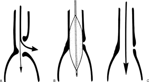
Figure 6.
Schematic drawing of balloon dilation of anastomotic leak. (A) When there is a leak at the anastomotic suture or staple line, esophageal secretions are forced to escape chiefly through the leak (large curved arrow) as flow into the distal gut is hampered by the narrowing of the anastomosis. (B) Balloon dilation of the anastomosis increases its diameter, thus (C) facilitating the flow of secretions into the distal gut and promoting closure and healing of the anastomotic leak. (Reprinted with permission from de Lange EE, Shaffer HA Jr, Holt PD. Esophagoenteric anastomotic leaks: treatment with fluoroscopically guided balloon dilatation. AJR Am J Roentgenol 1994;162:51–54, figure 2.)

