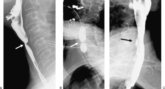
Figure 7.
A 59-year-old man with tight stricture of the cervical esophagus following laryngectomy. (A) Preprocedural esophagogram showing the stricture in the cervical esophagus. (B) The stricture was dilated with a 20-mm-diameter balloon. There is a waist corresponding to the stricture site (arrow). (C) An small intramural tear is demonstrated on the postprocedural esophagogram as a collection of extraluminal contrast material confined within the esophageal wall (arrow).

