Figure 1.
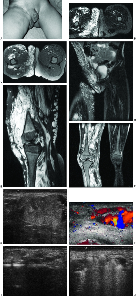
Diffuse venous malformation (VM) of the right lower extremity and perineum. This young girl became symptomatic before one year of age and has undergone numerous therapeutic procedures including sclerotherapy, resection of intraarticular VM of the right knee, and contour resection of the right labia majora. (A) Clinical photograph demonstrates enlargement of the right labia majora and adjacent buttock with purplish discoloration. Note the prominent varicosities in the adjacent thigh and pelvic wall. (B) T2-weighted axial magnetic resonance image (MRI) of the perineum shows diffuse involvement of the perineum with serpiginous, T2-hyperintense venous channels. (C) T2-weighted axial MRI of the perineum one year later, after several sessions of sclerotherapy and contour resection. Although the contour is improved, there is still VM throughout the labium and buttock. (D) Coronal T2-weighted MRI, 5 years after (B), and following contour resection of the right labium majorum shows a more symmetrical perineum. Note the involvement of the wall of the right pelvis, which has also been treated. (E) Sagittal T2-weighted MRI of the right knee, prior to treatment, showing a massive VM filling the joint and surrounding tissues. (F) Coronal T2-weighted MRI of the lower extremities after sclerotherapy and excision of the intraarticular VM. Note the severe swelling of the right lower extremity compared with the left. (G) Ultrasonographic image of the labium majorum in another patient with a focal VM of the perineum. The lesion is composed of tubular spaces that are compressible. Note the echogenic contents of the abnormal venous spaces and the fluid levels. (H) Color Doppler ultrasonogram shows blood flow or movement with release of compression by the ultrasound probe. (I) Ultrasonographic image demonstrating placement of a 16-gauge angiocatheter into the abnormal venous spaces. The metal stylette has been removed and a bare laser fiber inserted through the cannula. (J) Sonographic image during endovenous laser treatment. The steam generated by the laser energy causes echogenicity within the VM.

