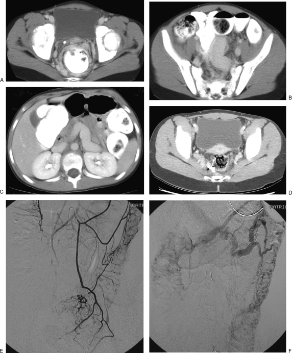
Figure 2.
Venous malformation (VM) involving the colon. This young girl presented with hematochezia. Imaging showed massive enlargement of the inferior mesenteric and hemorrhoidal veins. The inferior mesenteric vein was embolized through a transhepatic portal vein approach to decrease venous reflux into the malformation. Her bleeding diminished. However, 6 years later the patient developed acute portal vein thrombosis and required urgent endovascular recanalization. She remains anticoagulated. (A) Axial computed tomography (CT) image of the pelvis following intravenous and oral contrast administration demonstrates dilated venous channels arranged circumferentially around the rectum. Note, the phlebolith on the right anterior wall. (B,C) Axial CT images in the mid pelvis and abdomen show marked enlargement of the inferior mesenteric and portal veins. (D) Axial CT image of the pelvis 4 years after embolization shows some improvement in the venous engorgement around the rectum. (E,F) Inferior mesenteric arteriography 6 years after embolization shows a diffuse VM of the colon. Note the absence of filling of the inferior mesenteric vein.

