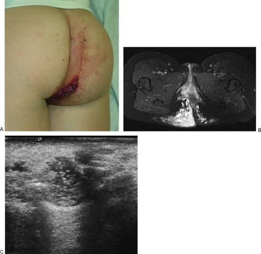
Figure 5.
Predominantly microcystic lymphatic malformation (LM) of the right buttock in an 18-year-old woman with recent onset of severe pain and leakage of bloody fluid from cutaneous vesicles. Resection of intraperitoneal cysts had been performed in the neonatal period. (A) The photograph demonstrates a capillary stain on the surface of the skin and an enlarged cluster of friable vesicles responsible for the leakage of serosanguineous fluid. (B) T2-weighted axial magnetic resonance image (MRI) shows increased thickness of the subcutaneous fat of the right buttock, with diffuse increased signal representing microcystic lymphatic malformation. Note the focal fluid collections that represent lymphatic cysts or spaces. The LM extends through the ischiorectal space into the retroperitoneum. (C) Ultrasonographic image during sclerotherapy shows microbubbles in one of the focal lymphatic spaces. This patient experienced dramatic improvement in pain and drainage following sclerotherapy.

