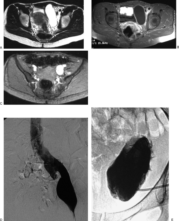
Figure 6.
Lymphaticovenous malformation (LVM) of the left pelvis in a 40-year-old woman with increasing pelvic fullness and discomfort. The symptoms improved after sclerotherapy of the lymphatic macrocysts. (A) T2-weighted axial magnetic resonance image (MRI) of the pelvis without fat suppression shows a large focal macrocyst to the left of the bladder and infiltration of the soft tissues around the iliac vessels with microcystic lymphatic malformation (LM). (B) Axial T1-weighted MRI after intravenous gadolinium and fat suppression shows enhancement of the rim of the macrocyst. (C) Gradient recalled echo image of the pelvis shows a large left iliac vein. (D) Left femoral venogram confirms the presence of dilatation of the left external iliac vein as well as the more proximal iliac veins and inferior vena cava. Dilatation of adjacent conducting veins is often seen in patients with macrocystic lymphatic malformations. (E) After placement of a pigtail catheter into the macrocyst, injection of contrast medium confirms the cystic nature of the malformation. Fluid was drained and the cyst was injected with sclerosant. Two procedures using ethanol as a sclerosant were ineffective. One treatment with OK-432 (picibanil) resulted in complete disappearance of the macrocyst.

