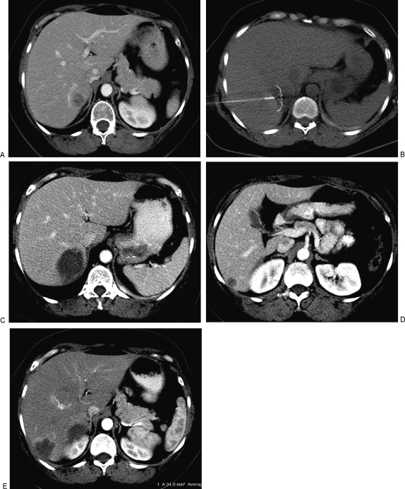
Figure 1.
(A) Contrast enhanced computed tomography (CT) image of a patient with metastatic breast cancer to bone and pleura that were well controlled; however, biopsy proved solitary metastasis in segment VI of the liver. (B) CT guided radiofrequency ablation (RFA) was performed with a Radio Therapeutics device shown with the tines deployed in the lesion. (C) CT scan 6 weeks after RFA demonstrates complete ablation of the target lesion with no residual hypervascular nodularity. (D) Three months after ablation of the lesion in (A), the patient had a second liver metastases, also in segment VI of the liver, which was treated with RFA. (E) Contrast enhanced CT 4 weeks after ablation of the second lesion demonstrates the typical smooth hypervascular rim of inflammatory tissue. Note the absence of hypervascular nodularity which would indicate residual viable tumor. Seven months after the first RFA, there is no evidence of viable liver metastases and the extrahepatic metastases to bone and pleura remain stable.

