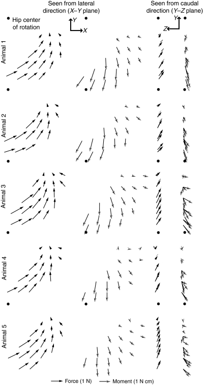
Fig. 3.
Force and moment fields evoked by in situ stimulation of the gracilis posticus (GRp) for each animal. First two columns show the recorded force (filled arrows) and moment (open arrows) field as seen from the lateral direction (X–Y plane; see Fig. 1A), and the last two columns show the field as seen from the caudal direction (Y–Z plane; see Fig. 1A). The position of the hip in each plot is indicated by the filled circle.

