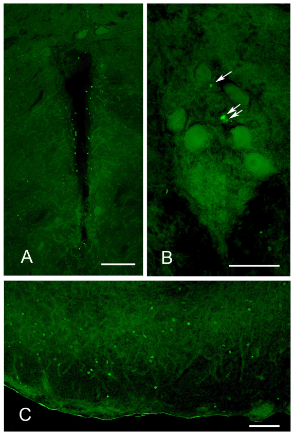
Fig. 2.
BrdU labeling in the brain of adult Brachyhypopomus gauderio. In all photographs, dorsal is towards the top of the figure. (A) Periventricular zone of the anterior midbrain. The cavity at the center is the ventricle. Scale bar, 100 μm. (B) Pacemaker nucleus. Arrows point to BrdU+ cells. The large cells are relay cells, which only show background labeling. Scale bar, 50 μm. (C) Electrosensory lateral line lobe (ELL). Figure shows one lateral half of the ELL, with lateral toward the left. Scale bar, 50 μm.

