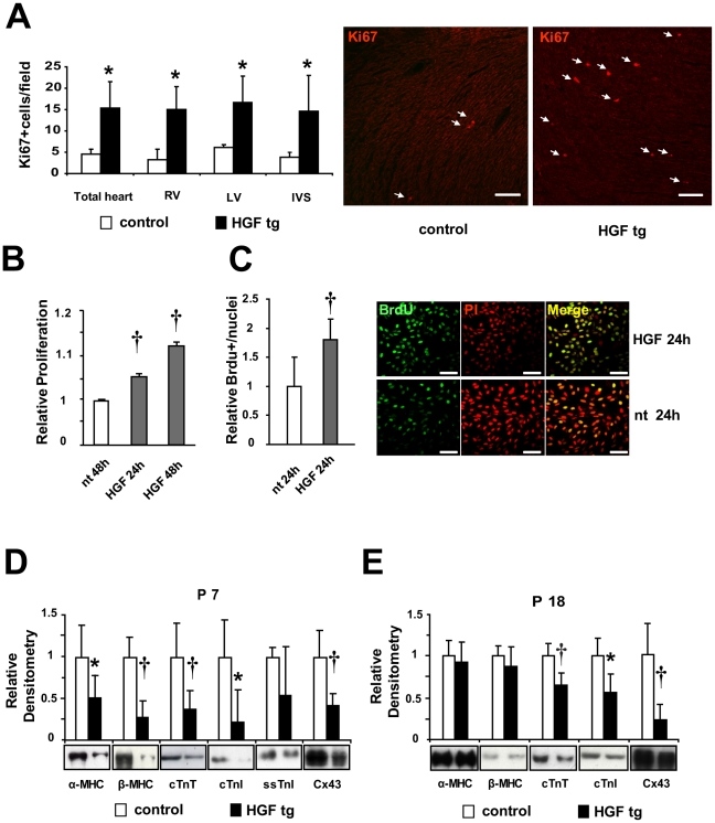
Figure 3. Increased proliferation and reduced expression of sarcomeric proteins and Connexin43 (Cx43) upon HGF induction in prenatal hearts.
(A) Left panel: Quantification of Ki67 positive nuclei in tissue sections of 7 days-old neonatal hearts. Right Ventricle (RV), Left Ventricle (LV) and Interventricular Septum (IVS) were separately or totally analyzed and compared in controls vs HGF tg neonates (n = 3 animals per group). At least 5 fields per zone per sample were counted. *p<0.05 (one-tailed T-test). Right panel: Representative Ki67 staining in tissue sections of 7 days-old neonatal hearts of control (left) and HGF tg (right). Ki67: red-nuclear (white arrows). Bar: 100 µm. (B) AlamarBlue assay and (C) BrdU incorporation of H9c2 cell line not treated (nt) and treated with 10U/ml HGF for the indicated times. Experiments were done in 8 (B) and 2 (C) biological replicates for each sample group. †p<0.005 versus nt (two-tailed T-test). Right panels: representative IF. BrdU: green-nuclear; propidium iodide (PI): red-nuclear. Bar: 75 µm. (D,E) Densitometric quantification normalized to Erk2 loading control and representative Western blots of the indicated proteins. Results represent averaged values for immunoblot analyses performed on heart lysates in (D) P7 neonatal controls (n = 10) vs HGF tg (n = 11) and (E) P18 young adult controls (n = 9) vs HGF tg (n = 14). Myosin heavy chains (α and β-MHC), troponins (cTnT, cTnI and ssTnI) and Cx43 have been quantified. *p<0.05, †p<0.005 (two-tailed T-test).

