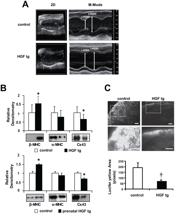
Figure 4. Contractile dysfunction, β-MHC re-expression and decreased Cx43 and cell-cell communication in adult HGF tg and prenatal HGF tg mice.
(A) Representative images of Left Ventricle long-axis echocardiogram (2D and M-Mode) of control (upper panels) and HGF tg mice (lower panels). (B) Densitometric quantification normalized to Erk2 loading control and representative Western blots of heart ventricles from control vs HGF tg (upper graph) n = 9 mice per group and prenatal HGF tg (lower graph) n = 3 mice per group. In the latter, HGF expression was suppressed after birth. Re-expression of β-MHC and decreased Cx43 are evident in both bitransgenic mice compared to controls. *p<0.05 (two-tailed T-test). (C) Representative images of Lucifer yellow dye diffusion in HGF tg and control edge-cut hearts (upper panels) and zoom-in of the areas included in dashed boxes (lower panels). Bottom graph: quantification of pixel area showed that cell-to-cell spread of Lucifer yellow was significantly decreased in HGF tg mice vs controls (n = 3 mice per group). †p<0.005 (one-tailed T-test). Bars: 100µm.

