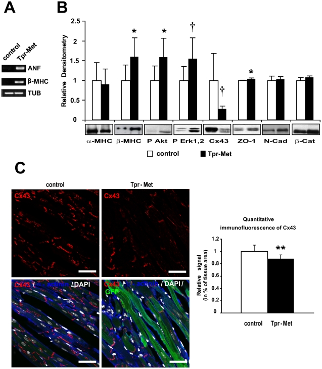
Figure 6. Tpr-Met expressing hearts show fetal gene re-expression and remodeling of Cx43.
(A) Semi-quantitative RT-PCR analysis of controls and postnatal Tpr-Met mice (P27) showed re-expression of ANF and β-MHC mRNA. Tubulin is used as loading control. (B) α to β isoform switch of MHC, increased phosphorylation of downstream Akt and Erk1,2 and strongly decreased Cx43, mild increase of ZO-1 and normal N-Cadherin and β-Catenin levels in postnatal Tpr-Met vs control hearts, analyzed by Western blot. Densitometric quantification normalized on Akt loading control and representative blots below graphs are shown. n = 10 mice for each group. *p<0.05 and †p<0.005 vs control (two-tailed T-test) (C) Left panels: representative confocal immunofluorescence images of left ventricle sections from postnatal Tpr-Met mice showed decreased staining of Cx43 (red), compared to controls (upper panels). Bottom panels: quadruple overlay with Cx43: red; α-actinin: blue; DAPI: white-nuclear; GFP: green-intracellular; Bars: 35µm. Quantification of Cx43 staining was performed with ImageJ. n = 6 controls and n = 3 postnatal Tpr-Met mice.

