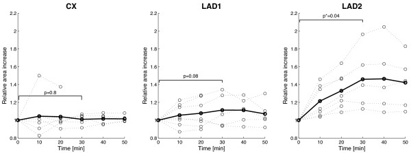
Figure 3.
Expansion of the contrast enhanced vessel area (CX, LAD1, and injured LAD2) as a function of time for all pigs (dotted lines) and average (bold lines). Thirty minutes post contrast, the injured LAD2 segment showed an average expansion of 45% (p = 0.04) while the average area of the uninjured CX and LAD1 artery remained constant with an average expansion of 7% (p = 0.8) and 11% (p = 0.08), respectively. In two pigs, images obtained 20 minutes post injection of gadofosveset were excluded due to poor image quality.

