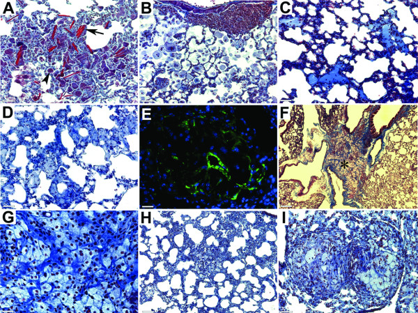
Figure 3.
Structural changes in lungs of Apoe-/-Ctsk-/- mice. (A), extensive fibrosis with Schaumann's bodies (arrowhead) and crystalline structures (arrow). (B), inducible bronchus-associated lymphoid tissues (iBALT) and alveolar monocyte/neutrophilic infiltration. (C), proteinosis. (D), ring-like structures formed by epithelioid cells. (E), smooth muscle cell α-actin positive cells in granulomas (green - smooth muscle cell α-actin, blue - nuclei). (F), accumulation of epithelioid cells between layers of collagen fibers (* shows area magnified on G). (H), individual epithelioid cells in fibrotic areas with honeycombing appearance. (I), two granulomas with relatively large size. (A-D, F-I), trichrome stained sections. (A-E and I), scale bars 65 μm; (F), scale bar 260 μm; (G), scale bar 30 μm; (H), scale bar 130 μm.

