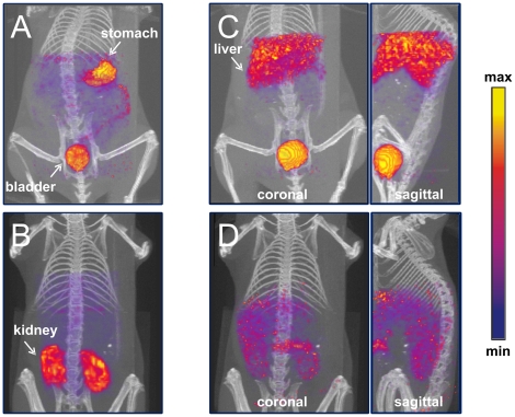
Figure 7. SPECT imaging analysis of a mouse injected with an adenovirus containing a capsid-incorporated pIX-MT fusion protein.
C57BL6 mice were injected intravenously with approximately 1 mCi of 99mTc in 0.3 mL PBS, and scanned at 30 min after the injection. Afterwards, 3D renderings of SPECT images in blue-purple-red-yellow scale against CT projections in grey scale were obtained. Shown are: (A) coronal image of a normal mouse injected with 99mTc-pertechnetate; (B) coronal image of a mouse injected with 99mTc-glucoheptonate; (C) coronal and sagittal images of a normal mouse injected with 99mTc-Ad-tGFP-pIX-MT; and (D) coronal and sagittal images of a mouse pretreated with warfarin injected with 99mTc-Ad-tGFP-pIX-MT.

