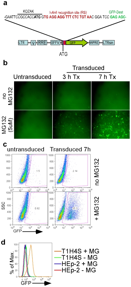
Figure 1. Cell line with integrated reporter lentivirus vector.
(a) Schematic of the reporter lentivirus. (b) Immunofluorescence imaging of cells 3 days post transduction (dpt) with reporter lentivirus, with or without MG132 for 3 or 7 h. (c) Flow cytometry of the cells from panel b after 7 h treatment with MG132. (d) Flow cytometry of the clonal reporter (T1H4S) or parental (HEp-2) cell line after 5 h incubation with (+ MG) or without (−MG) 1 µM MG132.

