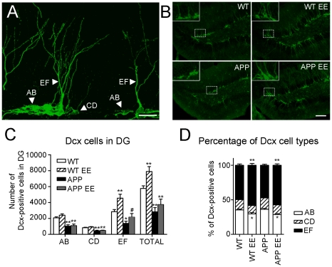
Figure 3. Environmental enrichment enhances the population of most-mature doublecortin cells in APPSw,Ind mice.
(A) Confocal microscopy image (maximum projection of several planes) of the dentate gyrus (DG) of an enriched WT mouse stained with doublecortin (Dcx) antibody showing the morphology of proliferative (type AB), intermediate (type CD) and postmitotic (type EF) neurons. Scale bar = 20 µm. (B) Representative epifluorescence microscopy images showing Dcx positive cells in the DG of the four experimental groups. Magnified images are shown in the insets. Scale bar = 100 µm. (C) Quantitative analysis reveals a significant reduction of total number of Dcx-labelled cells in the DG of APPSw,Ind mice (n = 6 mice) compared with WT mice (n = 6 mice). The number of EF-type neurons, but not AB- and CD-type neurons, is significantly increased in both WT (n = 6 mice) and APPSw,Ind mice (n = 6 mice) after EE. (D) Both enriched WT and APPSw,Ind mice show a significant increase in the percentage of EF cells and reduced percentage of AB cells compared to standard housed mice. Data represent mean ± SEM. *p<0.05 and **p<0.01, compared to WT mice. # p<0.05, compared to APP mice.

