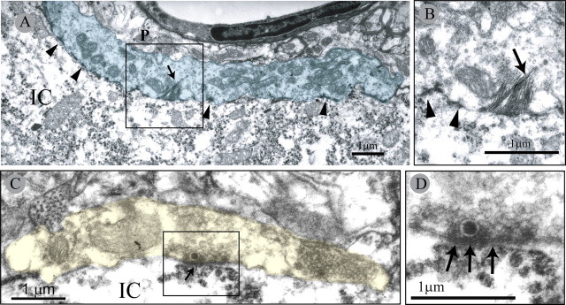
Figure 4.
Electron micrographs of M-type bouton apposing a γ-MN in a neonatally transected rat. A, High magnification of the M-bouton (blue). Note multiple active zones (arrowheads) and HRP product crystals (arrow). B, Detail of box in A showing HRP product crystals (arrow) and active sites (arrowheads). C, C-type bouton (yellow) apposing a γ-MN in a neonatally transected rat. D, Detail of subsynaptic cistern (arrows) associated with the postsynaptic element. Note the presence of a single large dense core vesicle near the presynaptic active zone region. IC, Motoneuron intracellular space; P, P-type bouton.

