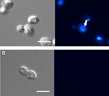
Figure 4.

DIC microscopy (left) and fluorescence microscopy with DAPI (right) of C. albicans (isolate 77) treated with 1 μg/ml of WSP1267 [IC50] for 48 h at 35ºC, showing abnormal chromatin condensation (A, white arrow) and absence of a nucleus (B). Bars = 5 μm.
