Figure 7.
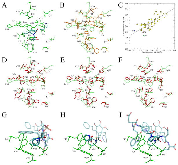
Binding site refinement for immunophilin FKBP12. (A) Binding pose of the FKB-001 ligand in the crystal structure of FKBP12 (PDB ID: 1j4r). FKB-001 is colored by atom type with the pipecolate moiety represented by thick sticks. (B) Binding pocket conformation in the structure modeled by chunk-TASSER (orange, solid) superposed onto the crystal structure (green, transparent). (C) Correlation between the observed and predicted RMSD from native for a non-redundant set of 50 binding site geometries constructed for FKBP12. Conformations at rank 1, 2 and 3 are colored in green, red and blue, respectively. (D, E and F) Top-ranked conformations (rank 1, 2 and 3, respectively) modeled by the BSR approach (red, solid) superimposed onto the crystal structure (green, transparent). (G, H and I) Ligands extracted from weakly related templates (PDB IDs: 2itk, 1pin and 2pv1, respectively) that contain conserved proline and pipecolate moieties (thick sticks colored by atom type) upon superposition of the template onto the target crystal structure. The anchor region is solid whereas the remaining part of the molecule is transparent. Thick (thin lines) indicate the ligand binding pose in the model (crystal structure). Selected interacting residues are shown in green.

