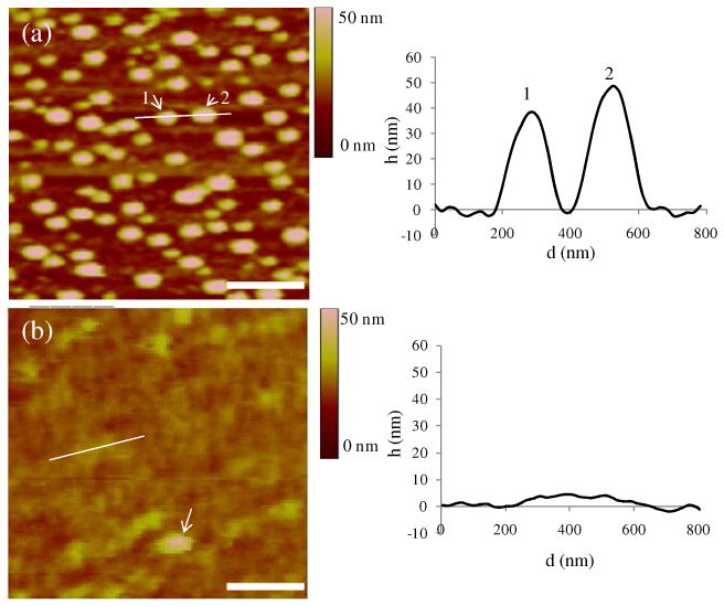
Figure 2.

AFM images of biotinylated vesicles captured on an avidin-functionalized surface after (a) rinsing in TBS and (b) rinsing in water. In (b), the line corresponds to the location of the measurement of the cross sectional height (right plot), and the arrow indicates the existence of an intact vesicle. (scale bar = 500 nm)
