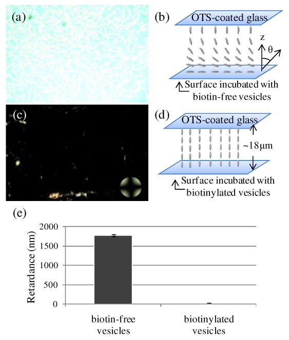
Figure 4.

(a) Optical image (crossed polars) of nematic 5CB sandwiched in an optical cell with an avidin-functionalized surface that was incubated against biotin-free vesicles. (b) Schematic illustration of the planar alignment of the LC in contact with the avidin-functionalized surface. The OTS-treated glass causes homeotropic anchoring of 5CB. (c) Optical image (crossed polars) of nematic 5CB sandwiched in an optical cell with an avidin-functionalized surface that was incubated against biotinylated vesicles. (d) Schematic illustration of the homeotropic anchoring of the LC on the surface incubated with biotinylated vesicles. (e) Optical retardance of the 5CB films described in (a) and (c). The error bars represent one standard deviation. (N=4)
