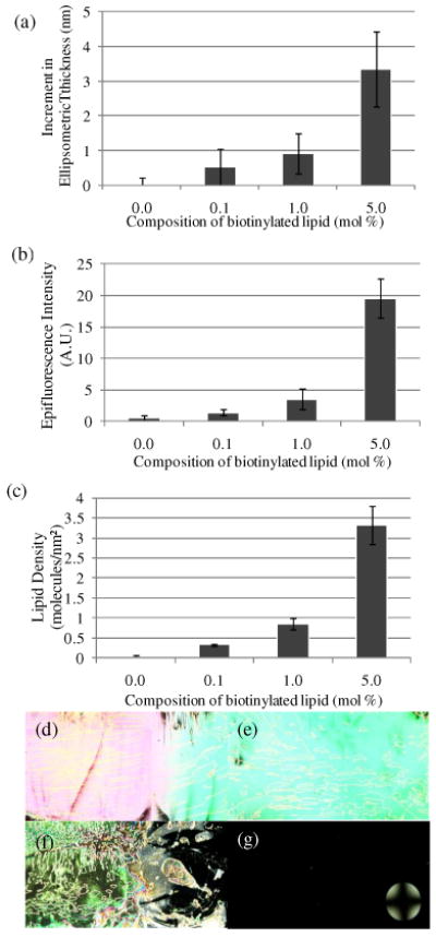
Figure 6.

(a) Increment in optical thickness following incubation of vesicles on avidin-functionalized surfaces, as a function of mole fraction of biotinylated lipid (0.1 mM total lipid concentration in solution). (b) Plot of epifluorescence intensity of surfaces measured following incubation of vesicles, as a function of mole fraction of biotinylated lipid. (c) Plot of lipid density measured following incubation of vesicles, as a function of mole fraction of biotinylated lipid. Optical images (crossed polars) of nematic 5CB sandwiched in optical cells with surfaces incubated with solutions of vesicles with (d) 0 mol %, (e) 0.1 mol %, (f) 1 mol % and (g) 5 mol % of biotinylated lipid. The error bars represent one standard deviation. (N=4)
