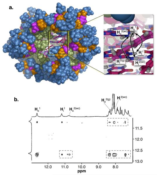
Figure 1.
(a) Molecular model of 316 constructed and minimized using MacroModel28 (the colors represent the different dendritic generations: green, D1; pink, D2; orange, D3; blue, D4). The inset shows a close up of the core of the assembly (carbons are shown in pink; oxygens in red; hydrogens in white; nitrogens in blue). The the double point arrows indicate to selected NOE interactions that give rise to the hexadecamer signature cross peaks. (b) 2D NOESY (500 MHz) showing a series of signature cross peaks that support the the structure of 316 (Figure S5).21,26

